

Veterinary anatomy reveals the inside secrets and beauty of the animal body
Virginia Maryland College of Veterinary Medicine, Department of Biomedical Sciences and Pathobiology
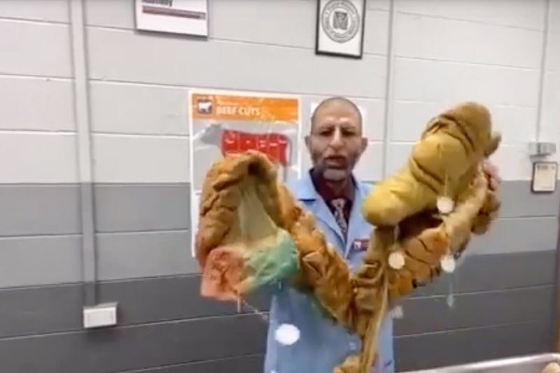
Anatomy uses ethically sourced cadavers preserved with formalin. Here, we display models preserved by an innovative method, the "Elnady Technique". I'm displaying the inside beauty of the animal body. Showing various real specimens preserved with Elnady technique. For example, stomachs of different domestic animals (horse, cow, calf, dog and cat) and we will explain why horses rarely vomit and why their stomach is different from us. We will display the chambers of the cow stomach and the interior of each chamber. The learner will see the large intestine of the horse and understand why horses frequently get colic. Sectioned horse’s head and distal limb and hoof will be displayed. I will also demonstrate some embryology stuff, such as fetuses of a goat, pig twins, and fetus of a cat. A pregnant uterus of a cow with an embryo inside showing how the placenta works and a goat fetus explaining how the blood circulates in the fetus to get his oxygen and nutrients from the mother and get rid of his waste products.
More exhibits
-
General Item
-
General Item
-
General Item
-
General Item
-
General Item


Page 1 of 8 | 47 Results